The objective of this study is to identify a clinical scenario for which normal ct derived right to left ventricular rv lv ratio.
Rv lv ratio pulmonary embolism.
Right ventricular dilatation rvd rv lv ratio 0 9 5.
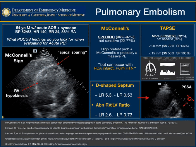
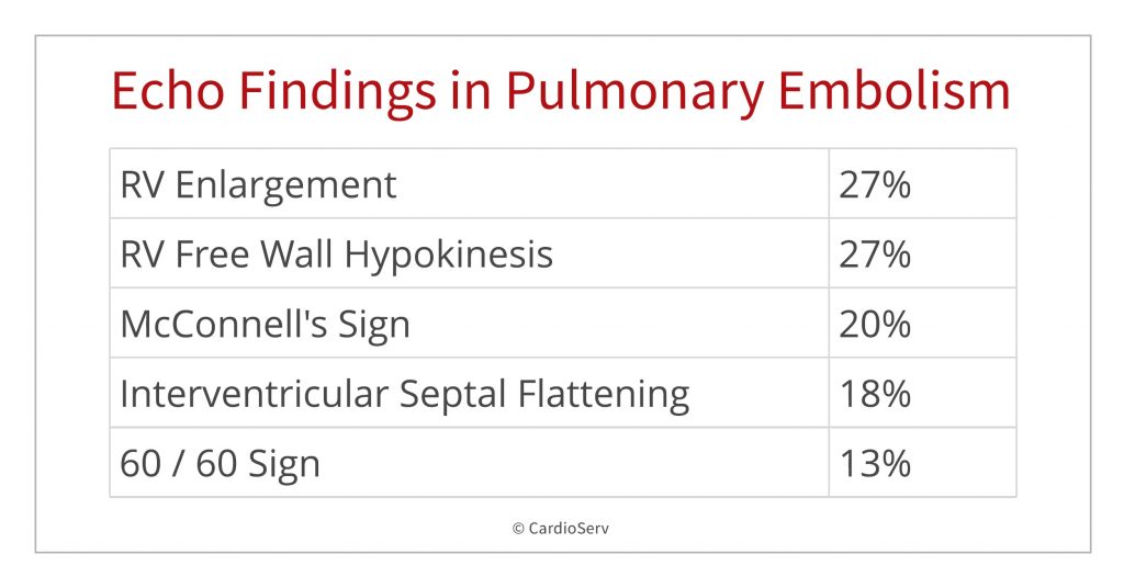
Mcconnell s sign 20.
Rv enlargement 27.
4 12 mg of tpa for 2 6 hrs.
Seattle ii submassive and massive pulmonary embolism treatment with ultrasound accelerated thrombolysis therapy 20.
10 major bleeding no ich.
The study assessed the frequency of echo findings in pulmonary embolism with the following findings.
May have a role in assessment.
Right ventricular wall can be thickened 4 mm often observed in congenital heart disease or dilated in acquired heart disease free wall may be hypokinetic.
Am j respir crit care med.
A right ventricle left ventricle rv lv ratio 1 0 was not associated with fewer favorable outcomes in patients with symptomatic acute pulmonary embolism pe who were otherwise considered low risk according to study results published in the american journal of respiratory and critical care medicine.
Plethoric inferior vena.
0 42 difference in rv lv ratio.
Epub ahead of print rv lv ratio measurement seems to have no role in low risk patients with pulmonary embolism treated at home triaged by hestia criteria.
Normal ventricular diameter ratio on ct provides adequate assessment for critical right ventricular strain among patients with acute pulmonary embolism the international journal of cardiovascular imaging 32 7 2016.
Rv free wall hypokinesis 27.
This is best appreciated on parasternal long axis projections.
There is variability in guideline recommendations for assessment of the right ventricle rv with imaging as prognostic information after acute pulmonary embolism pe.
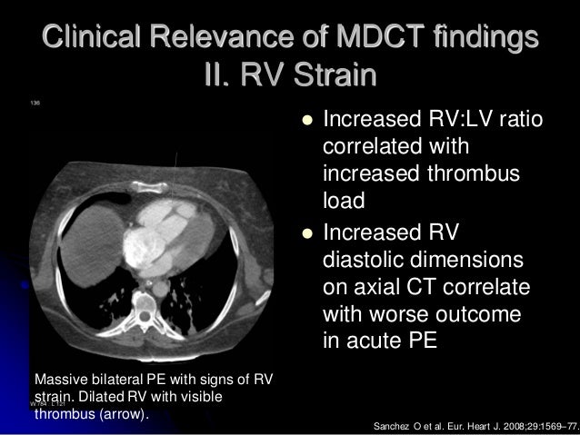
In the study by araoz et al 42 an rv lv diameter ratio greater than 1 was associated with a 3.
24 mg of tpa.
Demographic characteristics ild subtype echocardiography and.
Optalyse pe optimum duration of acoustic pulse thrombolysis procedure in acute pulmonary embolism 21.
Elevated right ventricular pressures.
Right ventricular systolic pressure 35 mmhg is consistent the 60 60 sign has gained recent attention putatively indicating an acute cause of elevated right ventricular pressures with a pulmonary valve acceleration time 60 ms and a tricuspid regurgitation jet 30 but 60 mmhg.
In this patient level post hoc analysis of 2 dutch clinical trials hestia.
0 3 0 4 difference in.
Additional studies have estimated that an rv lv diameter ratio superior to 1 5 indicates a severe episode of pe 36 39 41.
A jase study in 2016 analyzed the findings from 511 consecutive patients with pulmonary embolism.