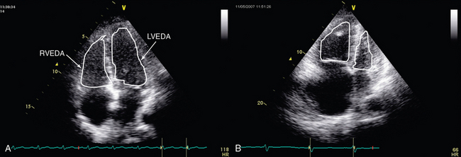
Normal 2d measurements from the apical 4 chamber view.
Rv lv ratio echocardiography.
Positive lr 29 and negative lr 0 51.
Rv medio lateral end diastolic dimension 4 3 cm rv end diastolic area 35 5 cm 2 maximal ra medio lateral and supero inferior dimensions 4 6 cm and 4 9 cm respectively maximal ra volume 33 ml m 2 35 89.
Used to demonstrate rv dilatation.
Sensitivity 50 specificity 98 ppv 88 npv 88.
Rv diastolic dysfunction 705 b.
It can manifest as an acute right heart syndrome.
Rv lv ratio echocardiogram lesson 174 part of our free online sonography training modules.
All patients with a mcconnell s sign were positive for pe.
Positive study rv dilation 1 1 ratio.
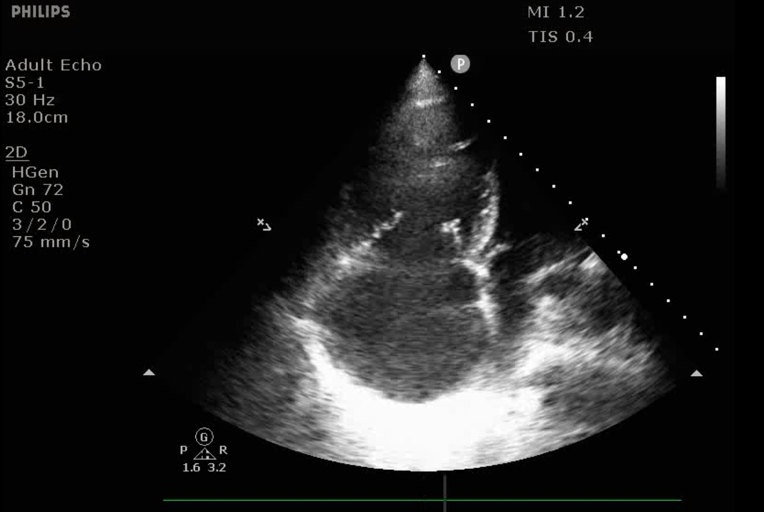
The assessment of rv function starts with the measurement of rv dimentions and the qualitative evaluation of its function.
The rv to left ventricular lv diameter rv lv ratio measured on ct imaging has been shown to predict the presence of ph in patients with pulmonary arterial hypertension 13 14 in addition studies have shown that the ct scan derived rv lv ratio predicts 30 day mortality in patients following acute pulmonary embolism.
17 patients with rv lv 1 1 and 15 found to have pe 2 false positives had copd 129 patients with no rv dilatation found to have pe 114 with no pe.
Echocardiography has variable sensitivity and specificity for the diagnosis of arvc and therefore.
Rv lv ratio 0 66 is abnormal a thickened or echo bright moderator band is not specific for arvc but may support the diagnosis in the presence of other find.
Effects of age respiration heart rate and loading condi tions 706.
Measurement of rv diastolic function 705 c.
Rv lv ratio measured on a positive study for acute pe is sensitive but highly nonspecific for all cause and pe related mortality high negative predictive value.
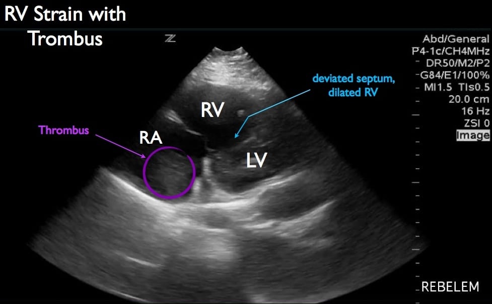
Right heart strain or more precisely right ventricular strain is a term given to denote the presence of right ventricular dysfunction usually in the absence of an underlying cardiomyopathy.
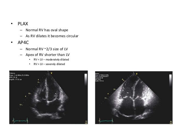
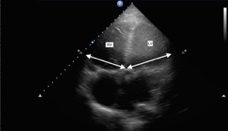
Notice the smaller rv surface compared to the lv aprox.
So normally the rv lv ratio should be about 0 6 to 1 and this is classically measured in the apical four chamber.
The right ventricle appears normal in size and systolic function.
The third e is for equality and this is really trying to estimate if the right ventricle is as big or bigger than the left ventricle.
To identify a clinical scenario for which ct rv lv ratio was considered sufficient to exclude rv strain or pe related short term death a multivariable logistic model was created to detect factors related to subjects for whom subsequent echocardiography detected rv strain or those who did not receive echocardiography and died of pe within 14.
While measurements are typically made using a true 4 chamber reformat made from the thin slice ct data.