The right ventricular to left ventricular diameter rv lv ratio measured at ct pulmonary angiogram ctpa has been shown to provide valuable information in patients with pulmonary arterial hypertension and to predict death or deterioration in acute.
Rv lv ratio echo.
ψ at a nyquist limit of 50 60 cm s.
To identify a clinical scenario for which ct rv lv ratio was considered sufficient to exclude rv strain or pe related short term death a multivariable logistic model was created to detect factors related to subjects for whom subsequent echocardiography detected rv strain or those who did not receive echocardiography and died of pe within 14.
Lv ei lv eccentricity index 1.
Sensitivity 50 specificity 98 ppv 88 npv 88.
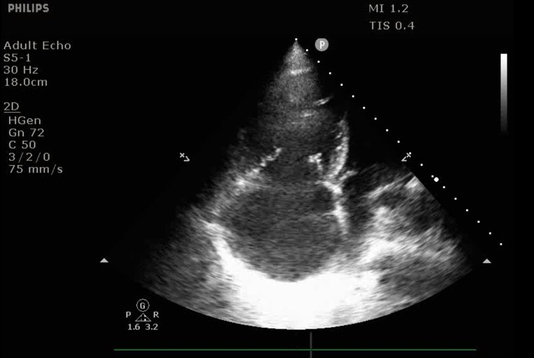
The right ventricle appears normal in size and systolic function.
In the absence of other etiologies of lv and la dilatation and acute mr.
Positive lr 29 and negative lr 0 51.
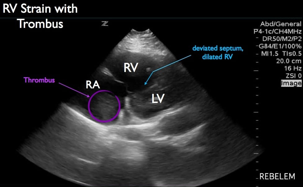
Rv lv ratio 0 9 rv strain ct pulmonary angiogram ctpa can not only visualize the clot but can also detect evidence of rv strain.
Right ventricle left ventricle end diastolic basal diameter ratio 1 the right ventricular outflow tract is considered enlarged when the measured diameter in the parasternal long axis exceeds 3 3 cm or when the measured diameter exceeds 2 7 cm in the distal rvot as measured in the basal parasternal short axis view.
17 patients with rv lv 1 1 and 15 found to have pe 2 false positives had copd 129 patients with no rv dilatation found to have pe 114 with no pe.
Rv lv ratio 0 66 is abnormal a thickened or echo bright moderator band is not specific for arvc but may support the diagnosis in the presence of other find ings there are no specific values for diagnosis of arvc however the measurement should be used to demonstrate ra dilatation.
All patients with a mcconnell s sign were positive for pe.
Rv medio lateral end diastolic dimension 4 3 cm rv end diastolic area 35 5 cm 2 maximal ra medio lateral and supero inferior dimensions 4 6 cm and 4 9 cm respectively maximal ra volume 33 ml m 2 35 89.
Patients with interstitial lung disease ild may develop pulmonary hypertension ph often disproportionate to the severity of the ild.
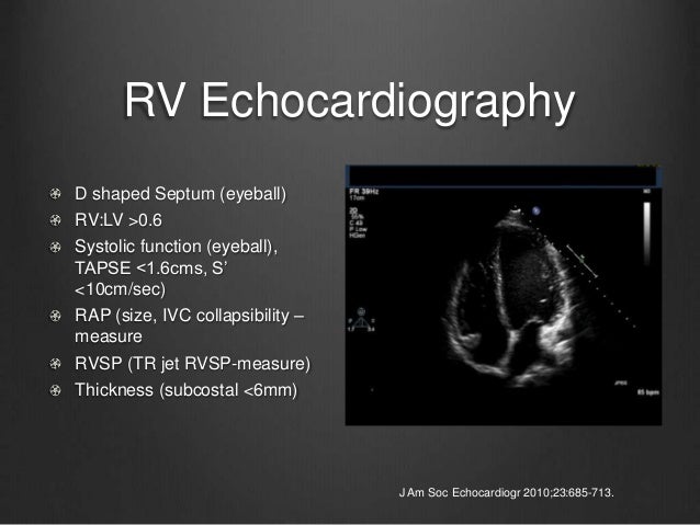
The assessment of rv function starts with the measurement of rv dimentions and the qualitative evaluation of its function.
Ra area 18cm2 is abnormal.
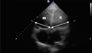
Normal 2d measurements from the apical 4 chamber view.
Regional assessment of rv systolic function 701 tapse or tricuspid annular motion tam 701 doppler tissue imaging 702 myocardial acceleration during isovolumic contraction 703 regional rv strain and strain rate 704 two dimensional strain 705.
Lv minor axis 2 8 cm m 2 lv end diastolic volume 82 ml m 2 maximal la antero posterior diameter 2 8 cm m 2 maximal la volume 36 ml m 2 2 33 35.
Used to demonstrate rv dilatation.
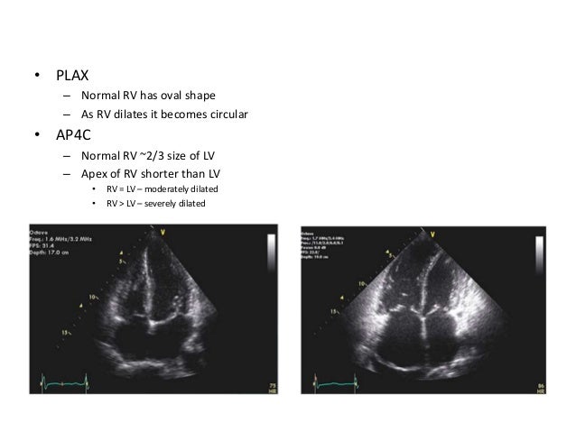
Notice the smaller rv surface compared to the lv aprox.